| NCBI National Center for Biotechnology Information |  |
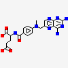
3EIG: Crystal structure of a methotrexate-resistant mutant of human dihydrofolate reductase |


Citing MMDB
 Madej T, Lanczycki CJ, Zhang D, Thiessen PA, Geer RC, Marchler-Bauer A, Bryant SH. " MMDB and VAST+: tracking structural similarities between macromolecular complexes. Nucleic Acids Res. 2014 Jan; 42(Database issue):D297-303