3K5V

Structure of Abl kinase in complex with imatinib and GNF-2


Experimental Data Snapshot

  • Method: X-RAY DIFFRACTION
  • Resolution: 1.74 Å
  • R-Value Free: 0.231 
  • R-Value Work: 0.199 
  • R-Value Observed: 0.201 

wwPDB Validation   3D Report Full Report


Ligand Structure Quality Assessment 


This is version 1.3 of the entry. See complete history


Literature

Targeting Bcr-Abl by combining allosteric with ATP-binding-site inhibitors.

Zhang, J.Adrian, F.J.Jahnke, W.Cowan-Jacob, S.W.Li, A.G.Iacob, R.E.Sim, T.Powers, J.Dierks, C.Sun, F.Guo, G.R.Ding, Q.Okram, B.Choi, Y.Wojciechowski, A.Deng, X.Liu, G.Fendrich, G.Strauss, A.Vajpai, N.Grzesiek, S.Tuntland, T.Liu, Y.Bursulaya, B.Azam, M.Manley, P.W.Engen, J.R.Daley, G.Q.Warmuth, M.Gray, N.S.

(2010) Nature 463: 501-506

  • DOI: https://doi.org/10.1038/nature08675
  • Primary Citation of Related Structures:  
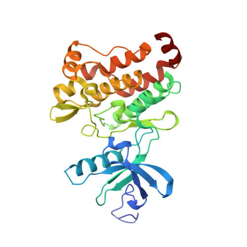
    3K5V

  • PubMed Abstract: 

    In an effort to find new pharmacological modalities to overcome resistance to ATP-binding-site inhibitors of Bcr-Abl, we recently reported the discovery of GNF-2, a selective allosteric Bcr-Abl inhibitor. Here, using solution NMR, X-ray crystallography, mutagenesis and hydrogen exchange mass spectrometry, we show that GNF-2 binds to the myristate-binding site of Abl, leading to changes in the structural dynamics of the ATP-binding site. GNF-5, an analogue of GNF-2 with improved pharmacokinetic properties, when used in combination with the ATP-competitive inhibitors imatinib or nilotinib, suppressed the emergence of resistance mutations in vitro, displayed additive inhibitory activity in biochemical and cellular assays against T315I mutant human Bcr-Abl and displayed in vivo efficacy against this recalcitrant mutant in a murine bone-marrow transplantation model. These results show that therapeutically relevant inhibition of Bcr-Abl activity can be achieved with inhibitors that bind to the myristate-binding site and that combining allosteric and ATP-competitive inhibitors can overcome resistance to either agent alone.


  • Organizational Affiliation

    Dana-Farber Cancer Institute, Harvard Medical School, Department of Cancer Biology, Seeley G. Mudd Building 628, Boston, Massachusetts 02115, USA.


Macromolecules
Find similar proteins by:  (by identity cutoff)  |  3D Structure
Entity ID: 1
MoleculeChains Sequence LengthOrganismDetailsImage
Tyrosine-protein kinase ABL1
A, B
293Mus musculusMutation(s): 0 
Gene Names: AblAbl1
EC: 2.7.10.2
UniProt
Find proteins for P00520 (Mus musculus)
Explore P00520 
Go to UniProtKB:  P00520
Entity Groups  
Sequence Clusters30% Identity50% Identity70% Identity90% Identity95% Identity100% Identity
UniProt GroupP00520
Sequence Annotations
Expand
  • Reference Sequence
Small Molecules
Binding Affinity Annotations 
IDSourceBinding Affinity
STI BindingDB:  3K5V Ki: min: 13, max: 7000 (nM) from 5 assay(s)
Kd: min: 1, max: 1.00e+4 (nM) from 35 assay(s)
IC50: min: 1.1, max: 1.00e+4 (nM) from 60 assay(s)
EC50: min: 34, max: 1000 (nM) from 4 assay(s)
STJ BindingDB:  3K5V Kd: 92 (nM) from 1 assay(s)
IC50: min: 9, max: 1.00e+4 (nM) from 4 assay(s)
EC50: 140 (nM) from 1 assay(s)
PDBBind:  3K5V Kd: 500 (nM) from 1 assay(s)
Experimental Data & Validation

Experimental Data

  • Method: X-RAY DIFFRACTION
  • Resolution: 1.74 Å
  • R-Value Free: 0.231 
  • R-Value Work: 0.199 
  • R-Value Observed: 0.201 
  • Space Group: P 1
Unit Cell:
Length ( Å )Angle ( ˚ )
a = 42.07α = 72.82
b = 65.268β = 80.25
c = 66.259γ = 84.86
Software Package:
Software NamePurpose
MAR345data collection
REFMACrefinement
XDSdata reduction
XSCALEdata scaling
REFMACphasing

Structure Validation

View Full Validation Report


Ligand Structure Quality Assessment 


Entry History 

Deposition Data

Revision History  (Full details and data files)

  • Version 1.0: 2010-01-19
    Type: Initial release
  • Version 1.1: 2011-07-13
    Changes: Version format compliance
  • Version 1.2: 2017-11-01
    Changes: Refinement description
  • Version 1.3: 2023-09-06
    Changes: Data collection, Database references, Derived calculations, Refinement description, Structure summary