6BT5

Human mGlu8 Receptor complexed with L-AP4


Experimental Data Snapshot

  • Method: X-RAY DIFFRACTION
  • Resolution: 2.92 Å
  • R-Value Free: 0.267 
  • R-Value Work: 0.253 
  • R-Value Observed: 0.254 

wwPDB Validation   3D Report Full Report


Ligand Structure Quality Assessment 


This is version 2.0 of the entry. See complete history


Literature

Determination of L-AP4-bound human mGlu8 receptor amino terminal domain structure and the molecular basis for L-AP4's group III mGlu receptor functional potency and selectivity.

Schkeryantz, J.M.Chen, Q.Ho, J.D.Atwell, S.Zhang, A.Vargas, M.C.Wang, J.Monn, J.A.Hao, J.

(2018) Bioorg Med Chem Lett 28: 612-617

  • DOI: https://doi.org/10.1016/j.bmcl.2018.01.037
  • Primary Citation of Related Structures:  
    6BSZ, 6BT5

  • PubMed Abstract: 

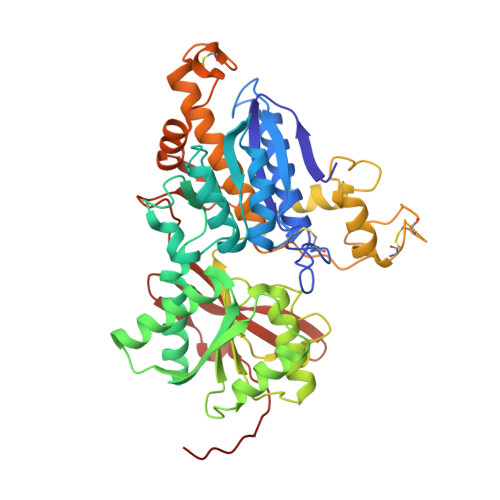
    L-2-Amino-4-phosphonobutyric acid (L-AP4) is a known potent and selective agonist for the Group III mGlu receptors. However, it does not show any selectivity among the individual group III mGlu subtypes. In order to understand the molecular basis for this group selectivity, we solved the first human mGlu8 amino terminal domain (ATD) crystal structures in complex with L-glu and L-AP4. In comparison with other published L-glu-bound mGlu ATD structures, we have observed L-glu binds in a significantly different manner in mGlu1. Furthermore, these new structures provided evidence that both the electronic and steric nature of the distal phosphate of L-AP4 contribute to its exquisite Group III functional agonist potency and selectivity.


  • Organizational Affiliation

    Discovery Chemistry Research and Technologies, Eli Lilly and Company, Lilly Corporate Center, Indianapolis, IN 46285, USA.


Macromolecules
Find similar proteins by:  (by identity cutoff)  |  3D Structure
Entity ID: 1
MoleculeChains Sequence LengthOrganismDetailsImage
Metabotropic glutamate receptor 8
A, B
479Homo sapiensMutation(s): 0 
Gene Names: GRM8GPRC1HMGLUR8
UniProt & NIH Common Fund Data Resources
Find proteins for O00222 (Homo sapiens)
Explore O00222 
Go to UniProtKB:  O00222
PHAROS:  O00222
GTEx:  ENSG00000179603 
Entity Groups  
Sequence Clusters30% Identity50% Identity70% Identity90% Identity95% Identity100% Identity
UniProt GroupO00222
Sequence Annotations
Expand
  • Reference Sequence
Small Molecules
Binding Affinity Annotations 
IDSourceBinding Affinity
E7P BindingDB:  6BT5 Ki: min: 61, max: 2700 (nM) from 5 assay(s)
IC50: 5.1 (nM) from 1 assay(s)
EC50: min: 60, max: 560 (nM) from 10 assay(s)
Experimental Data & Validation

Experimental Data

  • Method: X-RAY DIFFRACTION
  • Resolution: 2.92 Å
  • R-Value Free: 0.267 
  • R-Value Work: 0.253 
  • R-Value Observed: 0.254 
  • Space Group: I 2 2 2
Unit Cell:
Length ( Å )Angle ( ˚ )
a = 113.65α = 90
b = 166.008β = 90
c = 196.038γ = 90
Software Package:
Software NamePurpose
REFMACrefinement
PDB_EXTRACTdata extraction
XDSdata reduction
SCALAdata scaling
PHASERphasing

Structure Validation

View Full Validation Report


Ligand Structure Quality Assessment 


Entry History 

Deposition Data

Revision History  (Full details and data files)

  • Version 1.0: 2018-02-07
    Type: Initial release
  • Version 1.1: 2018-02-21
    Changes: Database references
  • Version 1.2: 2018-02-28
    Changes: Database references
  • Version 1.3: 2020-07-29
    Type: Remediation
    Reason: Carbohydrate remediation
    Changes: Data collection, Derived calculations, Structure summary
  • Version 2.0: 2023-11-15
    Changes: Atomic model, Data collection, Database references, Structure summary