3NC9

X-ray structure of ketohexokinase complexed with an indazole compound


Experimental Data Snapshot

  • Method: X-RAY DIFFRACTION
  • Resolution: 2.40 Å
  • R-Value Free: 0.285 
  • R-Value Work: 0.237 
  • R-Value Observed: 0.240 

wwPDB Validation   3D Report Full Report


Ligand Structure Quality Assessment 


This is version 1.4 of the entry. See complete history


Literature

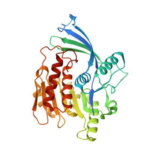
Electron density guided fragment-based lead discovery of ketohexokinase inhibitors.

Gibbs, A.C.Abad, M.C.Zhang, X.Tounge, B.A.Lewandowski, F.A.Struble, G.T.Sun, W.Sui, Z.Kuo, L.C.

(2010) J Med Chem 53: 7979-7991

  • DOI: https://doi.org/10.1021/jm100677s
  • Primary Citation of Related Structures:  
    3NBW, 3NC2, 3NC9, 3NCA

  • PubMed Abstract: 

    A fragment-based drug design paradigm has been successfully applied in the discovery of lead series of ketohexokinase inhibitors. The paradigm consists of three iterations of design, synthesis, and X-ray crystallographic screening to progress low molecular weight fragments to leadlike compounds. Applying electron density of fragments within the protein binding site as defined by X-ray crystallography, one can generate target specific leads without the use of affinity data. Our approach contrasts with most fragment-based drug design methodology where solution activity is a main design guide. Herein we describe the discovery of submicromolar ketohexokinase inhibitors with promising druglike properties.


  • Organizational Affiliation

    Johnson & Johnson Pharmaceutical Research and Development, Welsh and McKean Roads, Spring House, Pennsylvania 19477, USA.


Macromolecules
Find similar proteins by:  (by identity cutoff)  |  3D Structure
Entity ID: 1
MoleculeChains Sequence LengthOrganismDetailsImage
Ketohexokinase
A, B
313Homo sapiensMutation(s): 0 
Gene Names: KHK
EC: 2.7.1.3
UniProt & NIH Common Fund Data Resources
Find proteins for P50053 (Homo sapiens)
Explore P50053 
Go to UniProtKB:  P50053
PHAROS:  P50053
GTEx:  ENSG00000138030 
Entity Groups  
Sequence Clusters30% Identity50% Identity70% Identity90% Identity95% Identity100% Identity
UniProt GroupP50053
Sequence Annotations
Expand
  • Reference Sequence
Small Molecules
Binding Affinity Annotations 
IDSourceBinding Affinity
TR3 PDBBind:  3NC9 IC50: 330 (nM) from 1 assay(s)
Binding MOAD:  3NC9 IC50: 330 (nM) from 1 assay(s)
Experimental Data & Validation

Experimental Data

  • Method: X-RAY DIFFRACTION
  • Resolution: 2.40 Å
  • R-Value Free: 0.285 
  • R-Value Work: 0.237 
  • R-Value Observed: 0.240 
  • Space Group: P 21 21 21
Unit Cell:
Length ( Å )Angle ( ˚ )
a = 82.696α = 90
b = 85.692β = 90
c = 136.852γ = 90
Software Package:
Software NamePurpose
JDirectordata collection
PHENIXmodel building
PHENIXrefinement
d*TREKdata reduction
d*TREKdata scaling
PHENIXphasing

Structure Validation

View Full Validation Report


Ligand Structure Quality Assessment 


Entry History 

Deposition Data

Revision History  (Full details and data files)

  • Version 1.0: 2010-12-22
    Type: Initial release
  • Version 1.1: 2011-07-13
    Changes: Version format compliance
  • Version 1.2: 2017-02-22
    Changes: Structure summary
  • Version 1.3: 2017-11-08
    Changes: Advisory, Refinement description
  • Version 1.4: 2023-09-06
    Changes: Advisory, Data collection, Database references, Derived calculations, Refinement description