6D9H

Cryo-EM structure of the human adenosine A1 receptor-Gi2-protein complex bound to its endogenous agonist


Experimental Data Snapshot

  • Method: ELECTRON MICROSCOPY
  • Resolution: 3.60 Å
  • Aggregation State: PARTICLE 
  • Reconstruction Method: SINGLE PARTICLE 

wwPDB Validation   3D Report Full Report


This is version 1.2 of the entry. See complete history

Re-refinement Note

A newer entry is available that reflects an alternative modeling of the original data: 7LD4


Literature

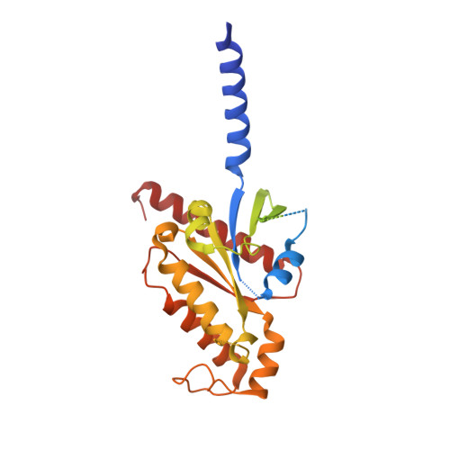
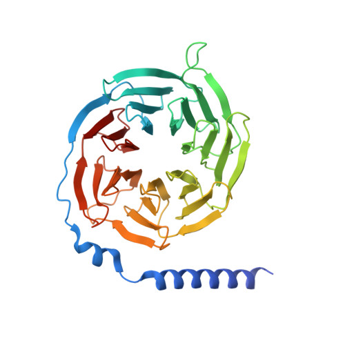
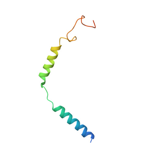
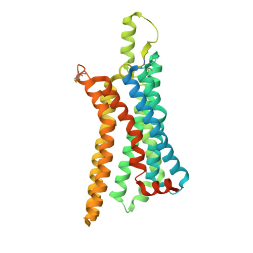
Structure of the adenosine-bound human adenosine A1receptor-Gicomplex.

Draper-Joyce, C.J.Khoshouei, M.Thal, D.M.Liang, Y.L.Nguyen, A.T.N.Furness, S.G.B.Venugopal, H.Baltos, J.A.Plitzko, J.M.Danev, R.Baumeister, W.May, L.T.Wootten, D.Sexton, P.M.Glukhova, A.Christopoulos, A.

(2018) Nature 558: 559-563

  • DOI: https://doi.org/10.1038/s41586-018-0236-6
  • Primary Citation of Related Structures:  
    6D9H

  • PubMed Abstract: 

    The class A adenosine A 1 receptor (A 1 R) is a G-protein-coupled receptor that preferentially couples to inhibitory G i/o heterotrimeric G proteins, has been implicated in numerous diseases, yet remains poorly targeted. Here we report the 3.6 Å structure of the human A 1 R in complex with adenosine and heterotrimeric G i2 protein determined by Volta phase plate cryo-electron microscopy. Compared to inactive A 1 R, there is contraction at the extracellular surface in the orthosteric binding site mediated via movement of transmembrane domains 1 and 2. At the intracellular surface, the G protein engages the A 1 R primarily via amino acids in the C terminus of the Gα i α5-helix, concomitant with a 10.5 Å outward movement of the A 1 R transmembrane domain 6. Comparison with the agonist-bound β 2 adrenergic receptor-G s -protein complex reveals distinct orientations for each G-protein subtype upon engagement with its receptor. This active A 1 R structure provides molecular insights into receptor and G-protein selectivity.


  • Organizational Affiliation

    Drug Discovery Biology and Department of Pharmacology, Monash Institute of Pharmaceutical Sciences, Monash University, Parkville, Victoria, Australia.


Macromolecules
Find similar proteins by:  (by identity cutoff)  |  3D Structure
Entity ID: 1
MoleculeChains Sequence LengthOrganismDetailsImage
Guanine nucleotide-binding protein G(i) subunit alpha-2355Homo sapiensMutation(s): 4 
Gene Names: GNAI2GNAI2B
Membrane Entity: Yes 
UniProt & NIH Common Fund Data Resources
Find proteins for P04899 (Homo sapiens)
Explore P04899 
Go to UniProtKB:  P04899
PHAROS:  P04899
GTEx:  ENSG00000114353 
Entity Groups  
Sequence Clusters30% Identity50% Identity70% Identity90% Identity95% Identity100% Identity
UniProt GroupP04899
Sequence Annotations
Expand
  • Reference Sequence
Find similar proteins by:  (by identity cutoff)  |  3D Structure
Entity ID: 2
MoleculeChains Sequence LengthOrganismDetailsImage
Guanine nucleotide-binding protein G(I)/G(S)/G(T) subunit beta-1350Homo sapiensMutation(s): 0 
Gene Names: GNB1
UniProt & NIH Common Fund Data Resources
Find proteins for P62873 (Homo sapiens)
Explore P62873 
Go to UniProtKB:  P62873
PHAROS:  P62873
GTEx:  ENSG00000078369 
Entity Groups  
Sequence Clusters30% Identity50% Identity70% Identity90% Identity95% Identity100% Identity
UniProt GroupP62873
Sequence Annotations
Expand
  • Reference Sequence
Find similar proteins by:  (by identity cutoff)  |  3D Structure
Entity ID: 3
MoleculeChains Sequence LengthOrganismDetailsImage
Guanine nucleotide-binding protein G(I)/G(S)/G(O) subunit gamma-2C [auth G]71Homo sapiensMutation(s): 0 
Gene Names: GNG2
Membrane Entity: Yes 
UniProt & NIH Common Fund Data Resources
Find proteins for P59768 (Homo sapiens)
Explore P59768 
Go to UniProtKB:  P59768
PHAROS:  P59768
GTEx:  ENSG00000186469 
Entity Groups  
Sequence Clusters30% Identity50% Identity70% Identity90% Identity95% Identity100% Identity
UniProt GroupP59768
Sequence Annotations
Expand
  • Reference Sequence
Find similar proteins by:  (by identity cutoff)  |  3D Structure
Entity ID: 4
MoleculeChains Sequence LengthOrganismDetailsImage
Chimera protein of Muscarinic acetylcholine receptor M4 and Adenosine receptor A1D [auth R]389Homo sapiensMutation(s): 0 
Gene Names: CHRM4ADORA1
Membrane Entity: Yes 
UniProt & NIH Common Fund Data Resources
Find proteins for P08173 (Homo sapiens)
Explore P08173 
Go to UniProtKB:  P08173
PHAROS:  P08173
GTEx:  ENSG00000180720 
Find proteins for P30542 (Homo sapiens)
Explore P30542 
Go to UniProtKB:  P30542
PHAROS:  P30542
GTEx:  ENSG00000163485 
Entity Groups  
Sequence Clusters30% Identity50% Identity70% Identity90% Identity95% Identity100% Identity
UniProt GroupsP30542P08173
Sequence Annotations
Expand
  • Reference Sequence
Small Molecules
Ligands 1 Unique
IDChains Name / Formula / InChI Key2D Diagram3D Interactions
ADN (Subject of Investigation/LOI)
Query on ADN

Download Ideal Coordinates CCD File 
E [auth R]ADENOSINE
C10 H13 N5 O4
OIRDTQYFTABQOQ-KQYNXXCUSA-N
Binding Affinity Annotations 
IDSourceBinding Affinity
ADN BindingDB:  6D9H Ki: min: 5.1, max: 2.90e+4 (nM) from 6 assay(s)
IC50: 2.3 (nM) from 1 assay(s)
EC50: min: 39, max: 3388 (nM) from 5 assay(s)
Experimental Data & Validation

Experimental Data

  • Method: ELECTRON MICROSCOPY
  • Resolution: 3.60 Å
  • Aggregation State: PARTICLE 
  • Reconstruction Method: SINGLE PARTICLE 

Structure Validation

View Full Validation Report


Entry History 

Revision History  (Full details and data files)

  • Version 1.0: 2018-06-20
    Type: Initial release
  • Version 1.1: 2018-07-04
    Changes: Data collection, Database references
  • Version 1.2: 2018-07-11
    Changes: Data collection, Database references