5OHB

Cereblon isoform 4 from Magnetospirillum gryphiswaldense in complex with Piperidin-2-one (Valerolactam)


Experimental Data Snapshot

  • Method: X-RAY DIFFRACTION
  • Resolution: 1.70 Å
  • R-Value Free: 0.212 
  • R-Value Work: 0.169 
  • R-Value Observed: 0.171 

wwPDB Validation   3D Report Full Report


Ligand Structure Quality Assessment 


This is version 1.3 of the entry. See complete history


Literature

Chemical Ligand Space of Cereblon.

Boichenko, I.Bar, K.Deiss, S.Heim, C.Albrecht, R.Lupas, A.N.Hernandez Alvarez, B.Hartmann, M.D.

(2018) ACS Omega 3: 11163-11171

  • DOI: https://doi.org/10.1021/acsomega.8b00959
  • Primary Citation of Related Structures:  
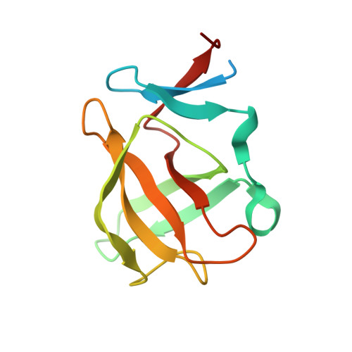
    5OH1, 5OH2, 5OH3, 5OH4, 5OH7, 5OH8, 5OH9, 5OHA, 5OHB

  • PubMed Abstract: 

    The protein cereblon serves as a substrate receptor of a ubiquitin ligase complex that can be tuned toward different target proteins by cereblon-binding agents. This approach to targeted protein degradation is exploited in different clinical settings and has sparked the development of a growing number of thalidomide derivatives. Here, we probe the chemical space of cereblon binding beyond such derivatives and work out a simple set of chemical requirements, delineating the metaclass of cereblon effectors. We report co-crystal structures for a diverse set of compounds, including commonly used pharmaceuticals, but also find that already minimalistic cereblon-binding moieties might exert teratogenic effects in zebrafish. Our results may guide the design of a post-thalidomide generation of therapeutic cereblon effectors and provide a framework for the circumvention of unintended cereblon binding by negative design for future pharmaceuticals.


  • Organizational Affiliation

    Department of Protein Evolution, Max Planck Institute for Developmental Biology, Max-Planck-Ring 5, 72076 Tübingen, Germany.


Macromolecules
Find similar proteins by:  (by identity cutoff)  |  3D Structure
Entity ID: 1
MoleculeChains Sequence LengthOrganismDetailsImage
Cereblon isoform 4
A, B, C
125Magnetospirillum gryphiswaldense MSR-1Mutation(s): 0 
Gene Names: MGR_0879
UniProt
Find proteins for A4TVL0 (Magnetospirillum gryphiswaldense)
Explore A4TVL0 
Go to UniProtKB:  A4TVL0
Entity Groups  
Sequence Clusters30% Identity50% Identity70% Identity90% Identity95% Identity100% Identity
UniProt GroupA4TVL0
Sequence Annotations
Expand
  • Reference Sequence
Experimental Data & Validation

Experimental Data

  • Method: X-RAY DIFFRACTION
  • Resolution: 1.70 Å
  • R-Value Free: 0.212 
  • R-Value Work: 0.169 
  • R-Value Observed: 0.171 
  • Space Group: P 21 21 21
Unit Cell:
Length ( Å )Angle ( ˚ )
a = 56.517α = 90
b = 59.63β = 90
c = 88.557γ = 90
Software Package:
Software NamePurpose
REFMACrefinement
XDSdata reduction
XDSdata scaling

Structure Validation

View Full Validation Report


Ligand Structure Quality Assessment 


Entry History 

Deposition Data

Revision History  (Full details and data files)

  • Version 1.0: 2018-10-10
    Type: Initial release
  • Version 1.1: 2018-11-28
    Changes: Data collection, Database references, Structure summary
  • Version 1.2: 2019-09-11
    Changes: Data collection, Database references
  • Version 1.3: 2024-01-17
    Changes: Data collection, Database references, Refinement description