3Q92

X-ray Structure of ketohexokinase in complex with a pyrimidopyrimidine analog 1


Experimental Data Snapshot

  • Method: X-RAY DIFFRACTION
  • Resolution: 2.80 Å
  • R-Value Free: 0.263 
  • R-Value Work: 0.202 
  • R-Value Observed: 0.207 

wwPDB Validation   3D Report Full Report


Ligand Structure Quality Assessment 


This is version 1.3 of the entry. See complete history


Literature

Inhibitors of Ketohexokinase: Discovery of Pyrimidinopyrimidines with Specific Substitution that Complements the ATP-Binding Site.

Maryanoff, B.E.O'Neill, J.C.McComsey, D.F.Yabut, S.C.Luci, D.K.Jordan, A.D.Masucci, J.A.Jones, W.J.Abad, M.C.Gibbs, A.C.Petrounia, I.

(2011) ACS Med Chem Lett 2: 538-543

  • DOI: https://doi.org/10.1021/ml200070g
  • Primary Citation of Related Structures:  
    3Q92, 3QA2, 3QAI

  • PubMed Abstract: 

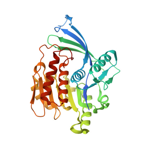
    Attenuation of fructose metabolism by the inhibition of ketohexokinase (KHK; fructokinase) should reduce body weight, free fatty acids, and triglycerides, thereby offering a novel approach to treat diabetes and obesity in response to modern diets. We have identified potent, selective inhibitors of human hepatic KHK within a series of pyrimidinopyrimidines (1). For example, 8, 38, and 47 exhibited KHK IC50 values of 12, 7, and 8 nM, respectively, and also showed potent cellular KHK inhibition (IC50 < 500 nM), which relates to their intrinsic potency vs KHK and their ability to penetrate cells. X-ray cocrystal structures of KHK complexes of 3, 8, and 47 revealed the important interactions within the enzyme's adenosine 5'-triphosphate (ATP)-binding pocket.


  • Organizational Affiliation

    Johnson & Johnson Pharmaceutical Research & Development , Spring House, Pennsylvania 19477-0776, United States.


Macromolecules
Find similar proteins by:  (by identity cutoff)  |  3D Structure
Entity ID: 1
MoleculeChains Sequence LengthOrganismDetailsImage
Ketohexokinase
A, B
313Homo sapiensMutation(s): 0 
Gene Names: KHK
EC: 2.7.1.3
UniProt & NIH Common Fund Data Resources
Find proteins for P50053 (Homo sapiens)
Explore P50053 
Go to UniProtKB:  P50053
PHAROS:  P50053
GTEx:  ENSG00000138030 
Entity Groups  
Sequence Clusters30% Identity50% Identity70% Identity90% Identity95% Identity100% Identity
UniProt GroupP50053
Sequence Annotations
Expand
  • Reference Sequence
Small Molecules
Binding Affinity Annotations 
IDSourceBinding Affinity
XNB PDBBind:  3Q92 IC50: 12 (nM) from 1 assay(s)
Experimental Data & Validation

Experimental Data

  • Method: X-RAY DIFFRACTION
  • Resolution: 2.80 Å
  • R-Value Free: 0.263 
  • R-Value Work: 0.202 
  • R-Value Observed: 0.207 
  • Space Group: P 21 21 21
Unit Cell:
Length ( Å )Angle ( ˚ )
a = 82.88α = 90
b = 86.073β = 90
c = 136.65γ = 90
Software Package:
Software NamePurpose
JDirectordata collection
PHENIXmodel building
PHENIXrefinement
d*TREKdata reduction
d*TREKdata scaling
PHENIXphasing

Structure Validation

View Full Validation Report


Ligand Structure Quality Assessment 


Entry History 

Deposition Data

  • Released Date: 2012-01-18 
  • Deposition Author(s): Abad, M.C.

Revision History  (Full details and data files)

  • Version 1.0: 2012-01-18
    Type: Initial release
  • Version 1.1: 2014-09-24
    Changes: Database references
  • Version 1.2: 2017-11-08
    Changes: Refinement description
  • Version 1.3: 2023-09-13
    Changes: Data collection, Database references, Derived calculations, Refinement description