4G7E

Crystal structure of pigeon pea urease


Experimental Data Snapshot

  • Method: X-RAY DIFFRACTION
  • Resolution: 2.20 Å
  • R-Value Free: 0.268 
  • R-Value Work: 0.217 
  • R-Value Observed: 0.220 

wwPDB Validation   3D Report Full Report


This is version 1.0 of the entry. See complete history


Literature

Structural and functional studies on urease from pigeon pea (Cajanus cajan)

Balasubramanian, A.Durairajpandian, V.Elumalai, S.Mathivanan, N.Munirajan, A.K.Ponnuraj, K.

(2013) Int J Biol Macromol 58C: 301-309

  • DOI: https://doi.org/10.1016/j.ijbiomac.2013.04.055
  • Primary Citation of Related Structures:  
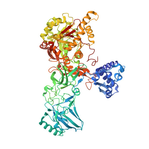
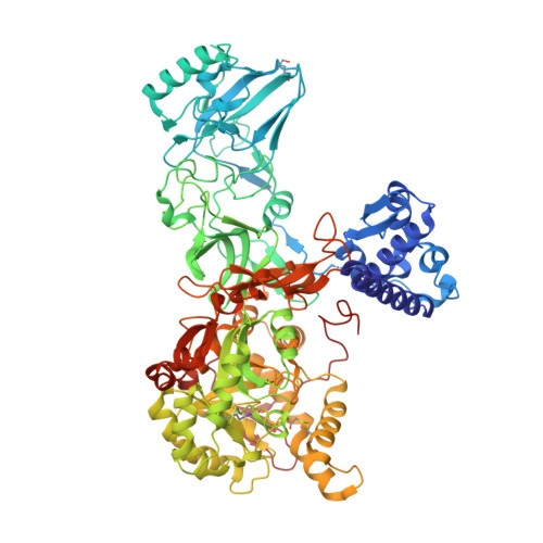
    4G7E

  • PubMed Abstract: 

    Urease is an enzyme that catalyzes the hydrolysis of urea, forming ammonia and carbon dioxide, and is found in plants, microorganisms and invertebrates. Although plant and bacterial ureases are closely related at amino acid and at the structural level, the insecticidal activity is seen only in the plant ureases. In contrast, both plant and bacterial ureases exhibit antifungal activity. These two biological properties are independent of its ureolytic activity. However, till date the mechanism(s) behind the insecticidal and fungicidal activity of ureases are not clearly understood. Here we report the crystal structure of pigeon pea urease (PPU, Cajanus cajan) which is the second structure from the plant source. We have deduced the amino acid sequence of PPU and also report here studies on its stability, insecticidal and antifungal activity. PPU exhibits cellulase activity. Based on the structural analysis of PPU and docking studies with cellopentoase we propose a possible mechanism of antifungal activity of urease.


  • Organizational Affiliation

    Centre of Advanced Study in Crystallography and Biophysics University of Madras, Guindy Campus, Chennai 600025, India.


Macromolecules
Find similar proteins by:  (by identity cutoff)  |  3D Structure
Entity ID: 1
MoleculeChains Sequence LengthOrganismDetailsImage
urease840Cajanus cajanMutation(s): 0 
EC: 3.5.1.5
UniProt
Find proteins for I3PA93 (Cajanus cajan)
Explore I3PA93 
Go to UniProtKB:  I3PA93
Entity Groups  
Sequence Clusters30% Identity50% Identity70% Identity90% Identity95% Identity100% Identity
UniProt GroupI3PA93
Sequence Annotations
Expand
  • Reference Sequence
Find similar proteins by:  (by identity cutoff)  |  3D Structure
Entity ID: 2
MoleculeChains Sequence LengthOrganismDetailsImage
urease840Cajanus cajanMutation(s): 0 
EC: 3.5.1.5
UniProt
Find proteins for I3PA93 (Cajanus cajan)
Explore I3PA93 
Go to UniProtKB:  I3PA93
Entity Groups  
Sequence Clusters30% Identity50% Identity70% Identity90% Identity95% Identity100% Identity
UniProt GroupI3PA93
Sequence Annotations
Expand
  • Reference Sequence
Experimental Data & Validation

Experimental Data

  • Method: X-RAY DIFFRACTION
  • Resolution: 2.20 Å
  • R-Value Free: 0.268 
  • R-Value Work: 0.217 
  • R-Value Observed: 0.220 
  • Space Group: H 3 2
Unit Cell:
Length ( Å )Angle ( ˚ )
a = 176.292α = 90
b = 176.292β = 90
c = 346.438γ = 120
Software Package:
Software NamePurpose
MAR345dtbdata collection
AMoREphasing
REFMACrefinement
AUTOMARdata reduction
SCALAdata scaling

Structure Validation

View Full Validation Report


Entry History 

Deposition Data

Revision History  (Full details and data files)

  • Version 1.0: 2013-06-05
    Type: Initial release