4NW7

PDE4 catalytic domain


Experimental Data Snapshot

  • Method: X-RAY DIFFRACTION
  • Resolution: 2.15 Å
  • R-Value Free: 0.207 
  • R-Value Work: 0.155 
  • R-Value Observed: 0.158 

wwPDB Validation   3D Report Full Report


Ligand Structure Quality Assessment 


This is version 1.1 of the entry. See complete history


Literature

Discovery of triazines as selective PDE4B versus PDE4D inhibitors.

Hagen, T.J.Mo, X.Burgin, A.B.Fox, D.Zhang, Z.Gurney, M.E.

(2014) Bioorg Med Chem Lett 24: 4031-4034

  • DOI: https://doi.org/10.1016/j.bmcl.2014.06.002
  • Primary Citation of Related Structures:  
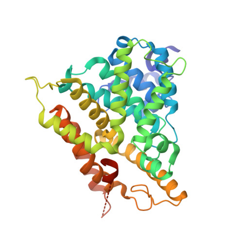
    4NW7

  • PubMed Abstract: 

    In this study we report a series of triazine derivatives that are potent inhibitors of PDE4B. We also provide a series of structure activity relationships that demonstrate the triazine core can be used to generate subtype selective inhibitors of PDE4B versus PDE4D. A high resolution co-crystal structure shows that the inhibitors interact with a C-terminal regulatory helix (CR3) locking the enzyme in an inactive 'closed' conformation. The results show that the compounds interact with both catalytic domain and CR3 residues. This provides the first structure-based approach to engineer PDE4B-selective inhibitors.


  • Organizational Affiliation

    Department of Chemistry and Biochemistry, Northern Illinois University, DeKalb, IL 60115, USA. Electronic address: thagen@niu.edu.


Macromolecules
Find similar proteins by:  (by identity cutoff)  |  3D Structure
Entity ID: 1
MoleculeChains Sequence LengthOrganismDetailsImage
cAMP-specific 3',5'-cyclic phosphodiesterase 4B372Homo sapiensMutation(s): 0 
Gene Names: DPDE4PDE4PDE4B
EC: 3.1.4.17
UniProt & NIH Common Fund Data Resources
Find proteins for Q07343 (Homo sapiens)
Explore Q07343 
Go to UniProtKB:  Q07343
PHAROS:  Q07343
GTEx:  ENSG00000184588 
Entity Groups  
Sequence Clusters30% Identity50% Identity70% Identity90% Identity95% Identity100% Identity
UniProt GroupQ07343
Sequence Annotations
Expand
  • Reference Sequence
Small Molecules
Ligands 5 Unique
IDChains Name / Formula / InChI Key2D Diagram3D Interactions
2O5
Query on 2O5

Download Ideal Coordinates CCD File 
E [auth A](4-{[4-(3-chlorophenyl)-6-cyclopropyl-1,3,5-triazin-2-yl]amino}phenyl)acetic acid
C20 H17 Cl N4 O2
CJQFUKZLNADPJB-UHFFFAOYSA-N
ZN
Query on ZN

Download Ideal Coordinates CCD File 
B [auth A]ZINC ION
Zn
PTFCDOFLOPIGGS-UHFFFAOYSA-N
EDO
Query on EDO

Download Ideal Coordinates CCD File 
F [auth A]
G [auth A]
H [auth A]
I [auth A]
J [auth A]
F [auth A],
G [auth A],
H [auth A],
I [auth A],
J [auth A],
K [auth A]
1,2-ETHANEDIOL
C2 H6 O2
LYCAIKOWRPUZTN-UHFFFAOYSA-N
MG
Query on MG

Download Ideal Coordinates CCD File 
C [auth A]MAGNESIUM ION
Mg
JLVVSXFLKOJNIY-UHFFFAOYSA-N
NA
Query on NA

Download Ideal Coordinates CCD File 
D [auth A]SODIUM ION
Na
FKNQFGJONOIPTF-UHFFFAOYSA-N
Binding Affinity Annotations 
IDSourceBinding Affinity
2O5 BindingDB:  4NW7 IC50: min: 237, max: 1181 (nM) from 2 assay(s)
Binding MOAD:  4NW7 IC50: 237 (nM) from 1 assay(s)
Experimental Data & Validation

Experimental Data

  • Method: X-RAY DIFFRACTION
  • Resolution: 2.15 Å
  • R-Value Free: 0.207 
  • R-Value Work: 0.155 
  • R-Value Observed: 0.158 
  • Space Group: P 42
Unit Cell:
Length ( Å )Angle ( ˚ )
a = 98.58α = 90
b = 98.58β = 90
c = 47.57γ = 90
Software Package:
Software NamePurpose
MD2data collection
PHASERphasing
REFMACrefinement
XDSdata reduction
XSCALEdata scaling

Structure Validation

View Full Validation Report


Ligand Structure Quality Assessment 


Entry History 

Deposition Data

Revision History  (Full details and data files)

  • Version 1.0: 2014-10-15
    Type: Initial release
  • Version 1.1: 2024-02-28
    Changes: Data collection, Database references, Derived calculations