3CE5

A bimolecular parallel-stranded human telomeric quadruplex in complex with a 3,6,9-trisubstituted acridine molecule BRACO19


Experimental Data Snapshot

  • Method: X-RAY DIFFRACTION
  • Resolution: 2.50 Å
  • R-Value Free: 0.213 
  • R-Value Work: 0.183 
  • R-Value Observed: 0.184 

wwPDB Validation   3D Report Full Report


Ligand Structure Quality Assessment 


This is version 1.3 of the entry. See complete history


Literature

Structural basis of DNA quadruplex recognition by an acridine drug.

Campbell, N.H.Parkinson, G.N.Reszka, A.P.Neidle, S.

(2008) J Am Chem Soc 130: 6722-6724

  • DOI: https://doi.org/10.1021/ja8016973
  • Primary Citation of Related Structures:  
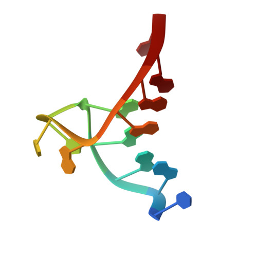
    3CE5

  • PubMed Abstract: 

    The crystal structure of a complex between the bimolecular human telomeric quadruplex d(TAGGGTTAGGGT)2 and the experimental anticancer drug BRACO-19, has been determined, to 2.5 A resolution. The binding site for the BRACO-19 molecule is at the interface of two parallel-folded quadruplexes, sandwiched between a G-tetrad surface and a TATA tetrad, and held in the site by networks of water molecules. The structure rationalizes the existing structure-activity data and provides a starting-point for the structure-based design of quadruplex-binding ligands


  • Organizational Affiliation

    Cancer Research UK Biomolecular Structure Group, The School of Pharmacy, University of London, 29-39 Brunswick Square, London WC1N 1AX, United Kingdom.


Macromolecules

Find similar nucleic acids by:  Sequence   |   3D Structure  

Entity ID: 1
MoleculeChains LengthOrganismImage
DNA (5'-D(*DTP*DAP*DGP*DGP*DGP*DTP*DTP*DAP*DGP*DGP*DGP*DT)-3')
A, B
12N/A
Sequence Annotations
Expand
  • Reference Sequence
Small Molecules
Ligands 2 Unique
IDChains Name / Formula / InChI Key2D Diagram3D Interactions
BRA
Query on BRA

Download Ideal Coordinates CCD File 
E [auth A]9-[4-(n,n-dimethylamino)phenylamino]-3,6-bis(3-pyrrolidinopropionamido) acridine
C35 H43 N7 O2
RKPYSYRMIXRZJT-UHFFFAOYSA-N
K
Query on K

Download Ideal Coordinates CCD File 
C [auth A],
D [auth A]
POTASSIUM ION
K
NPYPAHLBTDXSSS-UHFFFAOYSA-N
Experimental Data & Validation

Experimental Data

  • Method: X-RAY DIFFRACTION
  • Resolution: 2.50 Å
  • R-Value Free: 0.213 
  • R-Value Work: 0.183 
  • R-Value Observed: 0.184 
  • Space Group: I 4
Unit Cell:
Length ( Å )Angle ( ˚ )
a = 70.338α = 90
b = 70.338β = 90
c = 34.293γ = 90
Software Package:
Software NamePurpose
REFMACrefinement
CrystalCleardata collection
d*TREKdata reduction
d*TREKdata scaling
PHASERphasing

Structure Validation

View Full Validation Report


Ligand Structure Quality Assessment 


Entry History 

Deposition Data

Revision History  (Full details and data files)

  • Version 1.0: 2008-05-13
    Type: Initial release
  • Version 1.1: 2011-07-13
    Changes: Version format compliance
  • Version 1.2: 2017-01-11
    Changes: Other
  • Version 1.3: 2023-08-30
    Changes: Data collection, Database references, Derived calculations, Refinement description